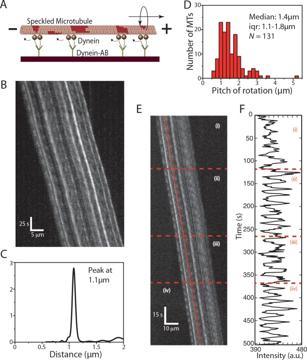
Fig 5. Cytoplasmic dynein driven MT rotations.
(A) S-MTs glide on a reflective silicon substrate coated with cytoplasmic dynein motor proteins specifically attached to the surface via anti-dynein antibodies. (B) Typical kymograph of a gliding S-MT. (C) Combined PSD curve obtained from nine speckles, with peak at about 1.1μm. (D) Histogram of the rotational pitches, median pitch of rotation is 1.4μm (iqr 1.1–1.8μm; N = 131, where N is the number of rotation events from 75 kymographs), which indicates that the gliding MTs rotate with shorter pitches than the supertwist of the employed GMP-CPP MTs. (E) Typical kymograph of a gliding S-MT with varying rotational pitch. The S-MT motion is divided into four sections as can be seen in (F), which shows the FLIC intensity versus time for one of the speckles on the MT lattice (indicated by the red line in E). Initially the MT had a rotational pitch of 1.1μm (i), then the rotational pitch reduced to 0.9μm (ii), followed by a switch to 1.1μm (iii), and finally, a reduction to 0.7μm (iv).

