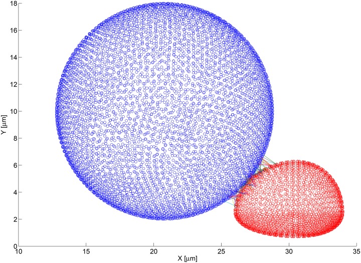
Fig 5. Bond formation between nearby cells using the fine computational mesh.
The blue figure is a 3D point cloud of the fine computational mesh used to describe the surface of the melanoma cell. Each blue circle represents the centroid of a discretized mesh face. Similarly, the red figure is a 3D point cloud of the fine computational mesh used to describe the surface of the PMN. The lines connecting the circles represents bonds that have formed and connect the two faces on which the involved adhesion molecules reside.

