
Figure 3.
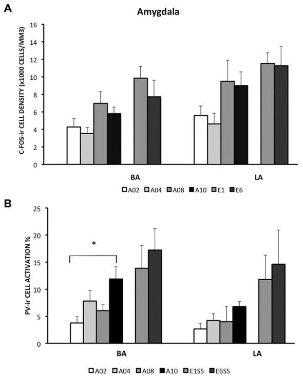
Densities of c-Fos-ir cells and the percent of activated PV-ir cells in the LA and BA were depicted. (A) Densities of c-Fos-ir cells in the lateral amygdala (LA) and basal amygdala (BA). (B) Activation of PV-ir cells in the LA and BA. Each data point represents group mean ± S.E.M. (*p < 0.05, n = 5–7/group).