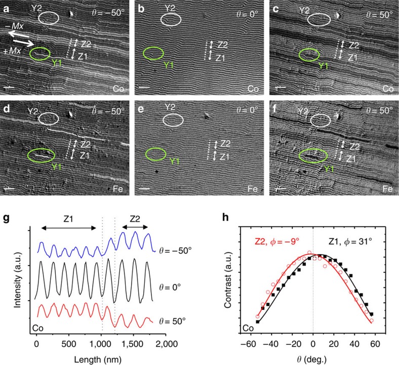
Figure 3. Characterization of in-plane and out-of-plane domain structure in NC/Py bilayer.
12 × 9 μm2 magnetic contrast images of the NdCo5/Py bilayer: Co magnetization at (a) θ=−50°, (b) θ=0° and (c) θ=+50°, and Fe magnetization at (d) θ=−50° , (e) θ=0° and (f) θ=+50°. Two typical dislocations denoted as Y1 (green ellipses) and Y2 (white ellipses) are shown. Scale bars, 1 μm. (g) Intensity profiles (Co edge) across dotted lines in panels (a–c). (h) Angular dependence of contrast curves at Z1 and Z2 regions (fitted φ=31° and −9°, respectively); corresponding Mx reversal is indicated by thick arrows in (a).

