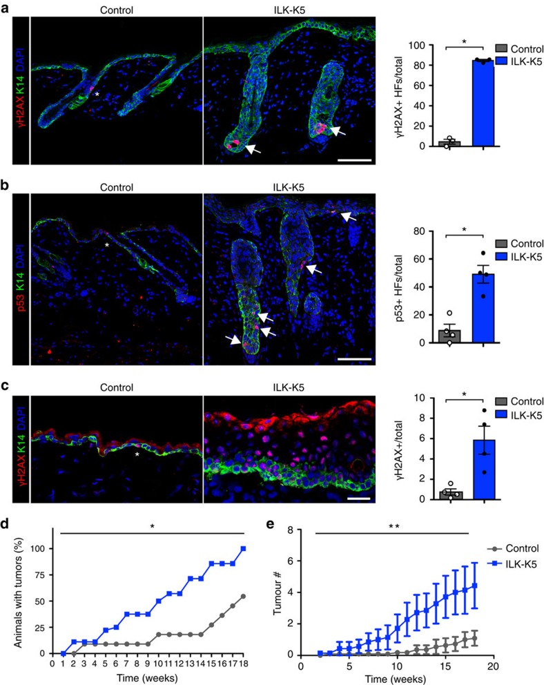
Figure 7. SC activation promotes replicative stress and skin carcinogenesis.
(a) Immmunofluorescence staining for γH2AX (red) and K14 (green) from P21 skin. Control mice rarely show γH2AX-positive cells (asterisk), whereas ILK-K5 HFs show clusters of cells with pan-nuclear γH2AX within HFs (arrows). Scale bars, 50 μm. Right panel shows quantification of HFs containing more than two γH2AX-positive cells (mean±s.e.m.; n=3; *P=0.0383, Mann–Whitney). (b) Staining for p53 (red) and K14 (green) from P21 skin. In contrast to control mice that show only solitary p53-positive cells (asterisk), ILK-K5 mice frequently show p53-positive cells within HFs and IFE (arrows). Scale bars, 50 μm. Right panel shows quantification of HFs containing more than two p53-positive cells (mean±s.e.m.; n=4; *P=0.0286, Mann–Whitney). (c) Staining for γH2AX (red) and K14 (green) from P57 skin. Control mice show only solitary γH2AX-positive cells (asterisk) within the IFE, whereas ILK-K5 IFE shows abundant pan-nuclear γH2AX staining. Scale bars, 50 μm. Right panel shows quantification of γH2AX-positive cells per total cells within the IFE (mean±s.e.m.; n=4; *P=0.0286, Mann–Whitney). (d) Tumour incidence of control and ILK-K5 mice treated twice with DMBA followed by 18 weeks of biweekly TPA treatment. ILK-K5 mice show increased tumour incidence (n=11/11; *P=0.0209, χ2-test). (e) Tumour multiplicity in affected control and ILK-K5 mice during 18 weeks of biweekly TPA treatment. ILK-K5 mice show increased tumour multiplicity (mean±s.e.m.; n=11/11; **P<0.01, two-way analysis of variance). DAPI, 4,6-diamidino-2-phenylindole.

