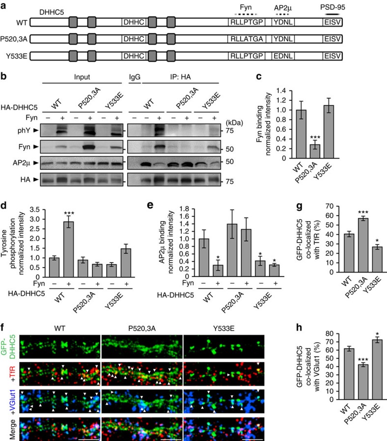
Figure 6. Phosphorylation of DHHC5 regulates its association with endocytic proteins and its subcellular localization.
(a) Schematic depiction of DHHC5 constructs N-terminally tagged with GFP or HA (not shown here) and illustrating the approximate localization of transmembrane domains (grey boxes), the DHHC motif, a putative Fyn-binding site (dashed line; RLLPTGP), a putative AP2μ-binding site (dashed line; YDNL) and the PDZ-binding motif required for binding PSD-95 (solid line; EISV). (b–e) HEK293T cells were transfected with the indicated HA-DHHC5 and Fyn constructs for 36 h, lysates immunoprecipitated with an HA antibody and blots probed with the indicated antibodies. (b,c) Fyn binding of the DHHC5 P520,3A mutant is reduced, but not for for the Y533E mutant (P=0.0153, F2,6=9.07). (b,d) Fyn-mediated tyrosine phosphorylation is attenuated in DHHC5 P520,3A and Y533E mutants (P<0.001, F5,12=20.04). (b,e) Fyn decreases AP2μ association with DHHC5 WT, but not P520,3A or Y533E mutants (P=0.018, F5,12=6.29). n=3 blots from 3 separate cultures. (f) Confocal images of 14 DIV neurons transfected with the indicated GFP–DHHC5 construct and immunostained for TfR and VGluT1. Scale bar, 5 μm. (g) DHHC5 P520,3A increases and Y533E decreases co-localization with TfR (P<0.001, F2,58=24.05), and (h) decreases and increases co-localization with VGlut1, respectively (P<0.001, F2,58=23.5). n=22 (WT), 18 (P520,3A) and 21 (Y533E) cells from 3 cultures. Co-localized puncta are denoted by white arrowheads. All graphs display mean±s.e.m. *P<0.05, **P<0.01, ***P<0.001; one-way analysis of variance; Tukey's post-hoc test. (b) Five per cent of whole-cell lysates were loaded as inputs. Full-length blots are presented in Supplementary Fig. 5.

