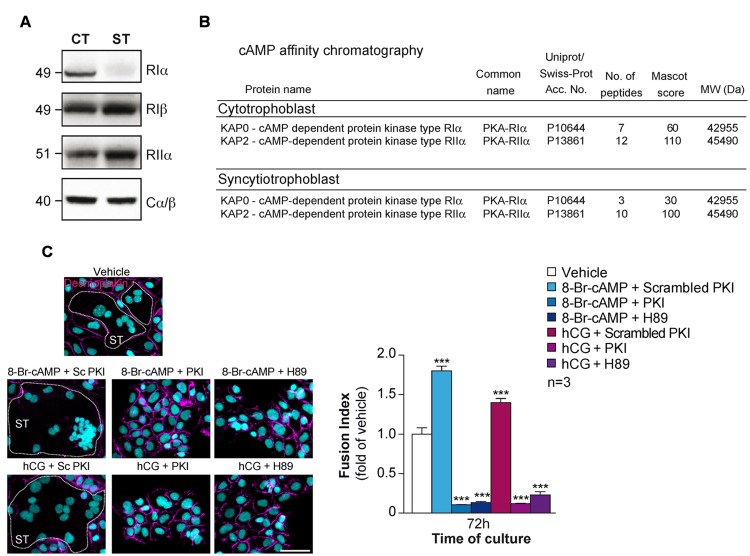
FIGURE 3.
Characterization of PKA in human trophoblasts. (A) Immunoblot analysis of RIα, RIβ, RIIα, and Cα/β in lysates of human primary trophoblasts at 24 and 72 h of culture. CT, ST (formed after 72 h of culture). (B) PKA R subunits identified by cAMP pulldown in trophoblasts. Proteins from CTs and STs were purified by cAMP affinity chromatography and identified by nanoLC-LTQ Orbitrap Mass Spectrometry analysis of tryptic digests of bands excised from SDS-PAGE. (C) Effect of 8-Br-cAMP (100 μM), hCG (1 μM), scrambled PKI or PKI peptide (10 μM each) and H89 (3 μM) on trophoblast fusion at 72 h of culture. Cells were immunostained for desmoplakin (magenta) and nuclei were counterstained with DAPI (left). Syncytia (ST) boundaries are indicated by dashed lines. Effect of 8-Br-cAMP, hCG in combination with PKI peptide or H89 on cell fusion represented as fusion indices histograms (upper right). Results are expressed as the mean ± SEM of n = 3 independent experiments (∗∗∗p < 0.001). Scale bar: 30 μm.

