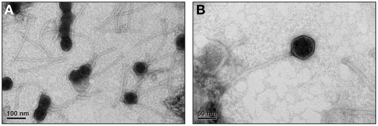
Figure 5.

Electron microgram of P1312 virions. The phage particles were prepared, negatively stained and examined by electron microscope as described in Materials and Methods. (A) The broad view of the phage particles. (B) The close-up of a single phage particle.
