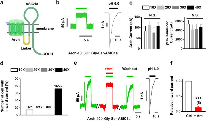
Figure 3. Characterization of Arch-ASIC1a interaction in single cells.
(a) Schematic diagram of Arch-ASIC1a chimera. A flexible linker (marked with yellow) of GS repeats is constituted by 10, 20, 30 or 40 amino acids. (b) Only Arch but not an ASIC-like current was observed when the linker was varied between 10 and 30 amino acids. The pH 6.0 (black bar)-induced current was used as a positive control. (c) Statistical results of light-activated Arch (left) and pH 6.0-induced ASIC1a currents (right) with various length of linker as indicated. Data represent means ± SEM (N.S., not significant of each two group, by unpaired t test, n = 7, 12, 8, 22 for 10, 20, 30 and 40 amino acids of linker, respectively). (d) The percentage of cells showing light-induced inward current in each group. (e,f), Light (530–550 nm filter, green bar)-induced ASIC1a activation by Arch only when the linker was 40 amino acids long (Arch-40 × Gly-Ser-ASIC1a). Ami, 100 μM (red bar). The pH 6.0 (black bar)-induced current was used as a positive control. (f) Summary data from (e) Ctrl, control. Data represent means ± SEM (***p < 0.001 by paired t test, n = 5).

