. 2015 Sep 15;4:e07068. doi: 10.7554/eLife.07068

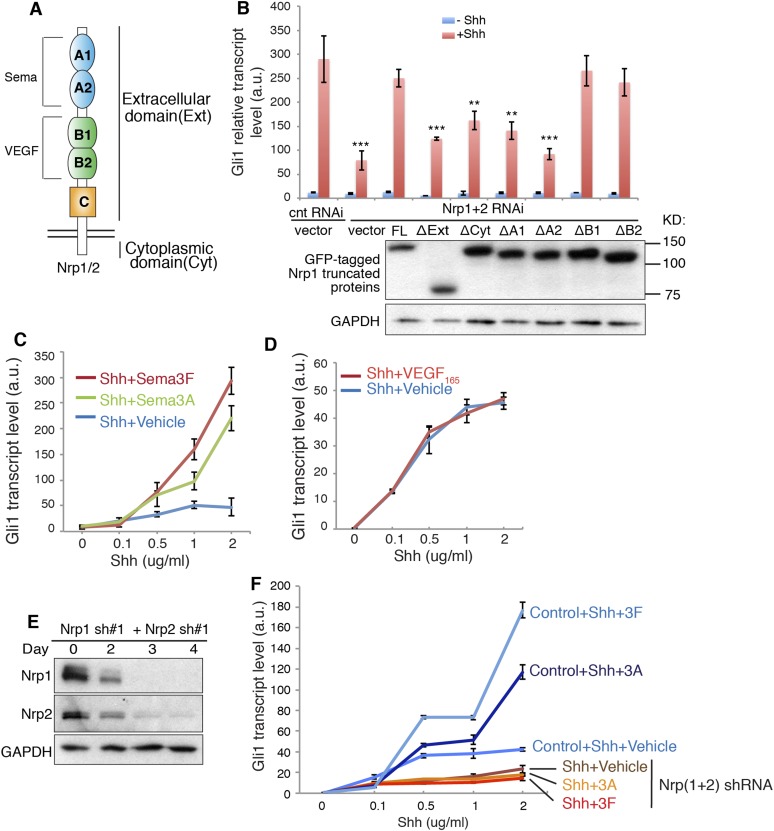
Figure 1. Signaling downstream of Sema3-Nrp enhances Hh transduction.

(A) Schematic drawing of Nrps protein structure. (B) In NIH3T3 cells in which Nrp1&2 were silenced by RNAi, Hh signaling could be rescued by full-length (FL) Nrp1 construct, but not by Nrp1 constructs that lack the entire extracellular domain (ΔExt), cytoplasmic domain (ΔCyt), A1 (ΔA1), or A2 (ΔA2) domain. Western blot shows that truncated Nrps were expressed with expected molecular weights. (C, D) Hh signaling activity in NIH3T3 cells treated with increasing concentrations of recombinant Shh in conjunction with a constant concentration of Sema3A, Sema3F (3 μg/ml), or VEGF165 (100 ng/ml) for 24 hr (E, F) Western blot shows that lentivirus-mediated expression of shRNA against Nrp1 and Nrp2 abolished the expression of endogenous Nrps (E). On day 3, Hh signaling activity was evaluated after cells were treated with Shh in conjunction with Sema3A or Sema3F for 24 hr (F). In all experiments Gli1 transcript level was measured by qPCR to evaluate Hh signaling activity; a.u., arbitrary unit. All error bars represent SEM. Statistics: Student's t-Test. *p < 0.05.

DOI: http://dx.doi.org/10.7554/eLife.07068.003

Figure 1.

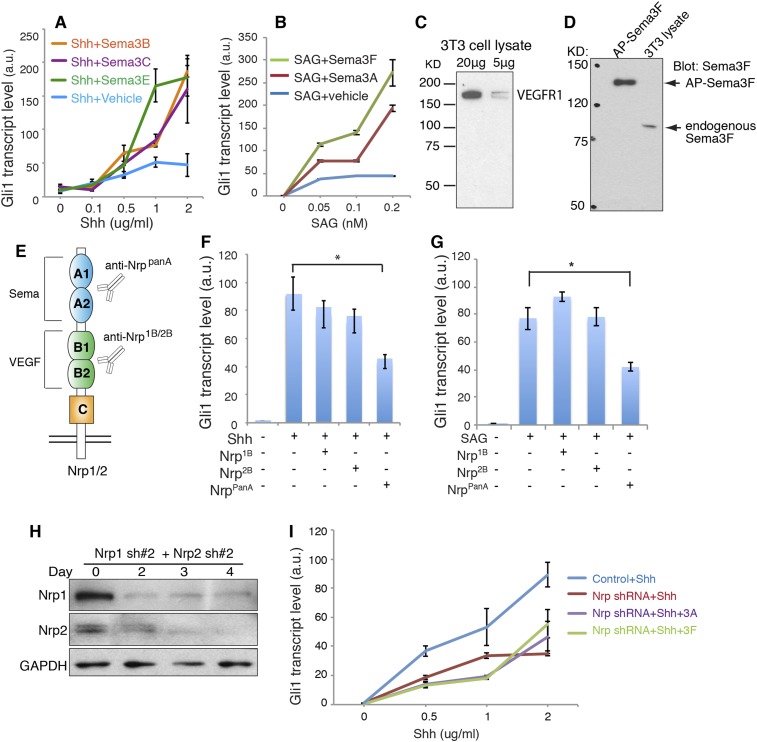
Figure 1—figure supplement 1. Sema3 signals through Nrps to enhance Hh transduction.

Figure 1—figure supplement 1.

(A, B) Hh signaling activity in NIH3T3 cells treated with increasing concentrations of Shh (A) or SAG (B) in conjunction with a constant concentration of Sema3 recombinant proteins (3 μg/ml) for 24 hr. (C, D) Western blot showing that VEGFR1 and Sema3F are expressed in 3T3 cells. (E) Diagram of function-blocking Nrp antibodies that specifically block Nrp binding with the ligands Sema3 and VEGF. (F, G) Hh signaling activity in NIH3T3 cells incubated with function-blocking Nrp antibodies (50 μg/ml) together with Shh or SAG for 4 hr. (H) Western blot shows that lentivirus-mediated expression of shRNA (pair #2) against Nrp1 and Nrp2 abolished the expression of endogenous Nrps. (I) On day 3, Hh signaling activity was evaluated after cells were treated with Shh in conjunction with Sema3A or Sema3F for 24 hr. In all experiments Gli1 transcript level was measured by qPCR to evaluate Hh signaling activity; a.u., arbitrary unit. All error bars represent SEM. Statistics: Student's t-Test. *p < 0.05, **p < 0.01, ***p < 0.001.