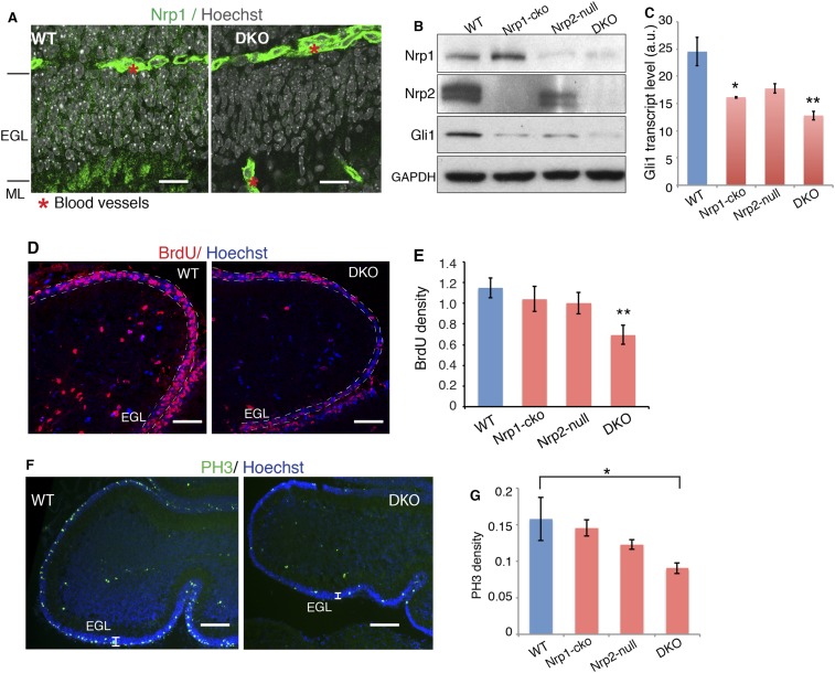
Figure 6. Loss of Nrps impairs GNP proliferation in the developing cerebellum.
(A) Immunostaining of Nrp1 (green) in the P7 cerebellum of wild type and Nrp1/2 DKO mouse show that Nrp1 was specifically knocked out from GNP cells in EGL and parallel fibers in ML, but remains in blood vessels. Scale, 20 μm. (B) Immunoblots showing that Nrp1 and 2 are knocked out from DKO cerebellum, and that Gli1 expression was much lower in the cerebellum of Nrp1/2 DKO mice compared to wild type littermates. Nrp1-cko: Math1Cre,Nrp1floxed/floxed. Nrp2-null: Nrp2-/-. (C) The Hh pathway activity in the cerebellum of Nrp1/2 DKO mice and littermates was evaluated by Gli1 transcript level. (D, E) GNP proliferation in the EGL (circled by dotted lines) of Nrp1/2 DKO mice and littermates was evaluated by BrdU incorporation assay. Scale: 50 μm. (F, G) Immunostaining and quantification of Phospho-H3 (green) in EGL of wild type and Nrp1/2 DKO cerebellum at P7. Scale, 100 μm. All error bars represent SEM. Statistics: non-parametric Mann–Whitney test. *p < 0.05, **p < 0.01.


