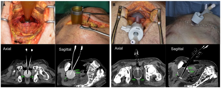
Figure 2.
Positioning of Zeiss Intrabeam™(A) or Xoft Axxent eBx™[(B) through laparoscopic trocar] applicators and CT scans of prostatectomized corpses with radiopaque clips located at different target tissues. U, urethra; ABN, anterior bladder neck; PBN, posterior bladder neck; LA, left apex; RA, right apex.

