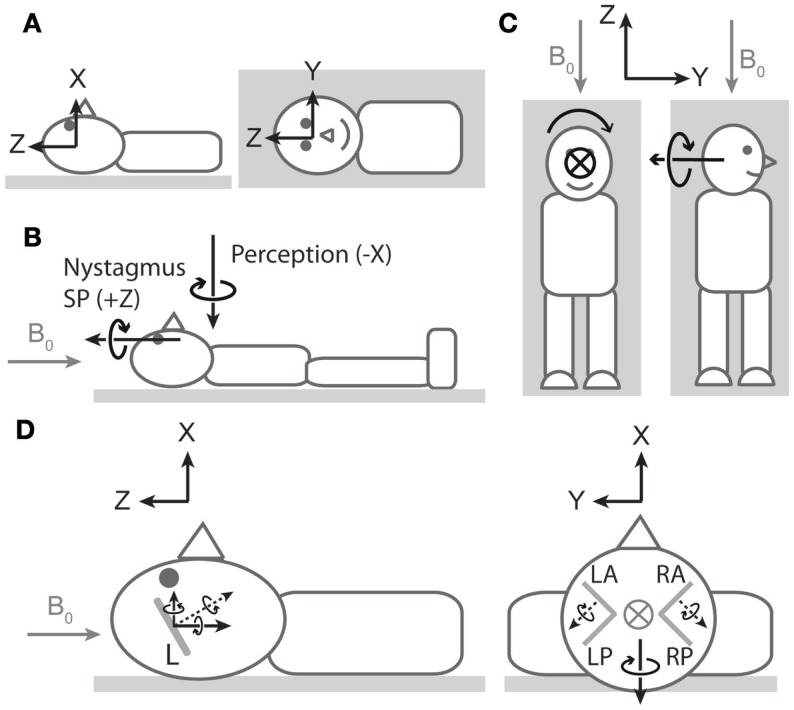
Figure 1.
Nystagmus, vertigo, and hypothetical canal signals during magnetic field exposure. In all cartoons, except the right side of (C), the head is in the neutral position described in the main text. All vectors are of arbitrary lengths and arbitrary relative lengths. Circle with cross denotes arrow entering page. Gray arrows denote magnetic field direction. (A) Coordinate system viewed from −Y (left cartoon) and +X (right cartoon) axes1. To describe direction of rotations, we use the right-hand grip rule (point thumb of right hand in direction of axis, and your fingers curl in the direction of positive rotation). Thus, a leftward eye rotation is +Z, and a perception of rotation in which the legs rotate to the right in the earth-horizontal plane is −X. (B) Vectors representing dominant components of nystagmus slow phase (SP) and rotation perception upon exposure to head-to-toe directed magnetic field are orthogonal. The nystagmus SP is +Z in most participants, and the perception of rotation is −X. The earth-horizontal location of the perception vector is arbitrary2. (C) Hypothetical rotation signal responsible for perception of rotation about X (left) should signal perception of rotation about Y when head orientation is altered by 90° about Z (right). (D) Hypothetical canal signals viewed from −Y (left) and +Z (right). Gray lines within the head are oversize representations of the approximate orientations of lateral (left cartoon) and vertical (right cartoon) semicircular canals (L = lateral, LA = left anterior, LP = left posterior, RA = right anterior, RP = right posterior). Dotted black arrows are rotation vectors from the canals inferred from nystagmus SP responses. Solid black arrows within the head are components of the vectors in the coordinate system. Left: horizontal eye movements with a leftward SP (+Z) imply a rotation vector from lateral canals with −Z component3. Since the lateral canals are inclined by approximately 20°–25° (14, 15), it also has a +X component. This is incongruent with the −X perception of rotation at this head position. Right: excitatory rotation signal from the left anterior canal and inhibitory signal from the right anterior canal [hypothesized in Ref. (9)]. The Y components of these signals would cancel, but the X components would sum leading to a −X signal that is directionally congruent with perception of rotation.

