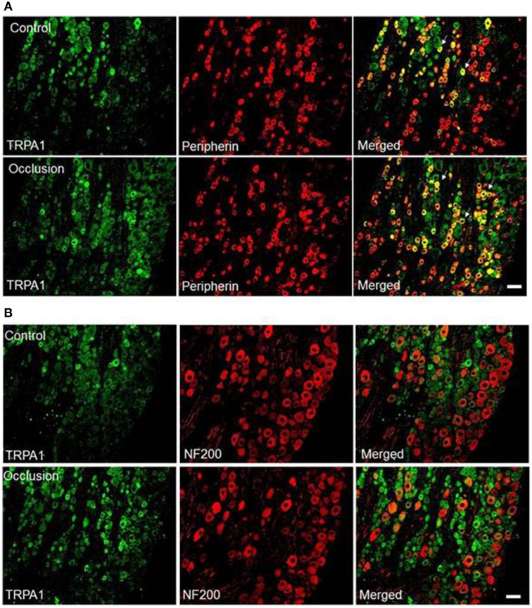
Figure 3.
Immunofluorescence was employed to examine double-labeling for TRPA1 and peripherin and NF200. Peripherin was used to label DRG neurons that supply thin C-fibers afferent nerves. NF200 was used to identify A-fibers of DRG neurons. (A) Representative photomicrographs show TRPA1 and peripherin staining in DRG neurons of a control limb (top) and a ligated limb (bottom). Arrows indicate representative cells positive for both TRPA1 and peripherin after they were merged. The number of double labeled DRG neurons is greater in ligated limbs than in control limbs. Scale bar = 50 μm. (B) Photomicrographs are representative to illustrate staining of TRPA1 and NF200 in DRG neurons of a control limb (top) and a ligated limb (bottom). There were few DRG neurons containing both TRPA1 and NF200 staining in both groups. No differences in the number of double-stained TRPA1 and NF200 were observed in DRG neurons of the control and ligated limbs. Scale bar = 50 μm.

