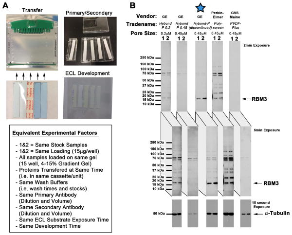
Fig. 1. Comparison of PVDF Membranes Used to Detect RBM3 by Western Blot.
(A) Methods used to equalize factors for comparison of PVDF membranes. Neuron homogenates (i.e. samples 1and 2) were loaded onto a 15-well 4–15% SDS gradient gel in five replicates. PVDF membranes were precisely cut to span the width of 2 lanes and match gel length. Proteins were transferred to all 5 membranes at the same time (in the same tank/cassette). Membranes were processed using the same volume/time incubation in blocking solution, TBS washes, primary antibody, secondary antibody, and ECL detection reagent. Membranes were put inside a single film holder for equivalent film exposures in a dark room. (B) Western blots show comparison of PVDF membranes to capture and/or detect RBM3.

