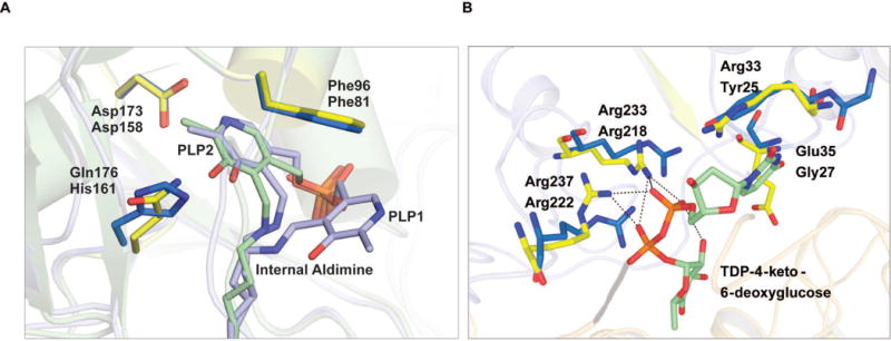
Figure 3.

Comparison of active site residues of AtmS13 with CalS13. (A) PLP binding site where the internal aldimine belonging to AtmS13 and CalS13 are colored blue and green, respectively. AtmS13 and CalS13 residues are colored marine blue and yellow, respectively. (B) Sugar nucleotide binding site (AtmS13 residues, marine blue; CalS13 residues, yellow; sugar nucleotide, green with pyrophosphate linkage in orange).
