Fig. 2.
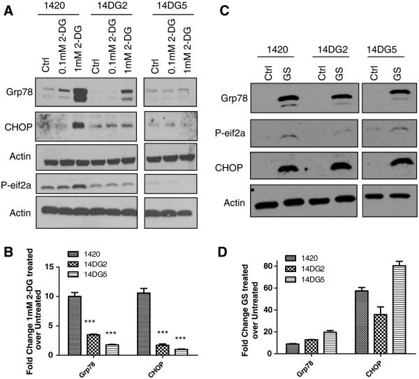
2-DG but not GS toxicity correlates with induction of UPR. a Cells were treated with the indicated doses of 2-DG for 24 h in normoxia and then harvested, and immunoblotting was performed to detect protein levels of Grp78, phospho-eif2a and CHOP. β-Actin was used as a loading control. b mRNA levels of Grp78 and CHOP were determined by qPCR in cells treated for 24 h with 1 mM 2-DG, normalized to β-actin and shown as fold induction of treated over untreated control samples. The bars represent the average of duplicate samples. ***P < 0.001 as compared with 1420. c Cells were treated with GS for 24 h in normoxia and then harvested, and immunoblotting was performed to detect protein levels of Grp78, phospho-eif2α and CHOP. β-Actin was used as a loading control. d mRNA levels of Grp78 and CHOP were determined by qPCR in cells treated for 24 h with GS and normalized to β-actin and shown as fold induction of treated over untreated control samples. The bars represent the average of duplicate samples

