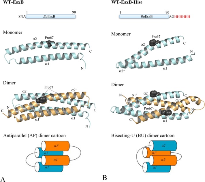
Figure 1.

The crystal structures and schematic diagrams of the antiparallel (AP, Panel A) and bisecting-U (BU, Panel B) topologies of EsxB. Top panels show the linear sequence and tags included in the recombinant proteins used for crystallization. Middle panels show the ribbon diagrams of the structure of EsxB monomers and dimers with Pro67 drawn in sphere representation to highlight the kink in α2. Monomers A and B are in pale cyan and light orange, respectively. The helical motifs within the inter-helices links of the BU dimer are labeled as α2° (see text for details). The lower panels show the schematic diagrams of the AP and BU dimers. Figures 1–4 are prepared with the program PyMOL (http://www.PyMOL.org).
