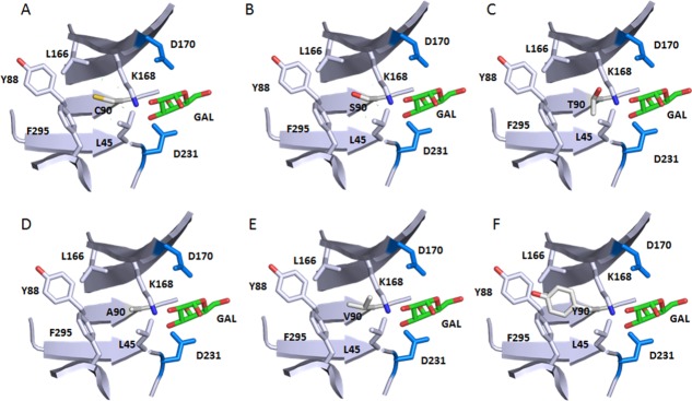
Figure 8.

Local environment of Cys90 and the effect of selective mutations. Only part of the backbone are shown for simplifications. The atom coloring scheme is same as in Figure 1 except for what is specified for each panel. Catalytic residues D170 and D231 and bound product GAL are shown too. Structure is from 3HG5.4 Residues surrounding residues of C90 form a hydrophobic pocket. (A) WT Cys90 sulfhydryl sulfur is shown in yellow. (B) C90S mutation with the Serine hydroxyl oxygen in red. (C) C90T mutation with the Threonine hydroxyl oxygen in red. (D) C90A. (E) C90V. (F) C90Y with the Tyrosine hydroxyl oxygen in red. C90Y clashes with nearby Y88.
