
Figure 1.
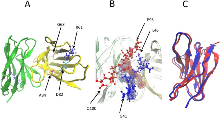
(A) Structure of dimer of the light-chain protein REI where yellow marks chain 1 and green does chain 2. The mutated residues are highlighted as blue = R61, red = G68, cyan = D82, and purple = A84. (B) The dimer interface of the REI light-chain protein. The second dimer interface is shown in the background, red = residues 94–101 of chain 1 and blue = residues 41–49 of chain 2. (C) Structural overlap of native and amyloid form of the REI light-chain protein.