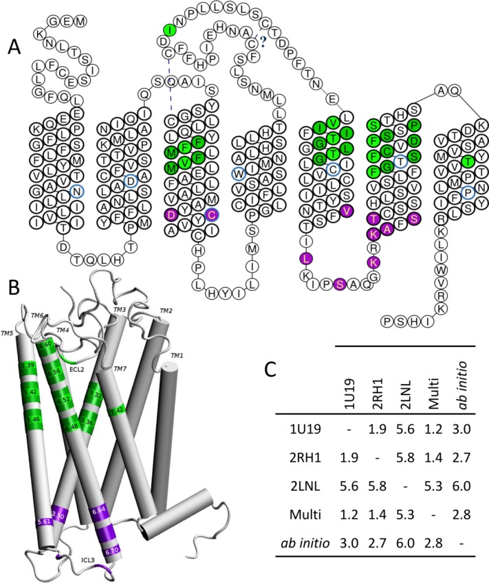
Figure 2.

Residues governing the function of mammalian ORs projected onto the sequence and the structure of hOR1G1. A, snakeplot of the OR sequence with residues involved in odorant contact in green and those involved in the OR activation through a contact with the G Protein in purple. Residues in light green will be strongly in contact with the odorant, those in dark green contribute to the wall of the binding cavity. Number 50 residue of the Ballesteros-Weinstein notation are circled in blue. The cysteine bridges are also indicated. B, position of important residues on the structure of the receptor, with some Ballesteros-Weinstein notations. C, C-α positions Root Mean Square deviation (in Å) between models build using Bovine Rhodopsin (PDB ID: 1U19), β2-adrenergic (PDB ID: 2RH1), Chemokine-1 (PDB ID: 2LNL) receptor, or a multitemplate (Multi) of the three receptors cited above, or an ab initio model (See Supporting Information for PDB structure of each model).
