
Fig. 1.
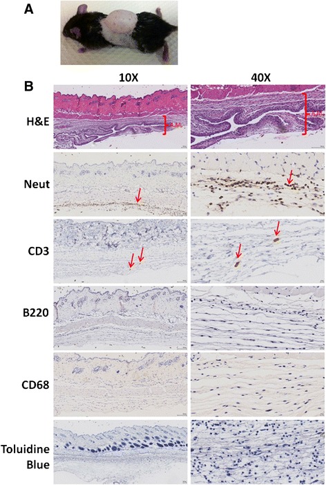
Air pouch model of inflammation. a Inflammation in the air pouch was induced as described. b H&E reveals the formation of the air pouch membrane (A.M.) and immunohistology staining with specific neutrophil (Neut), T cell (CD3), B cell (B220) and macrophage (CD68) markers, and the toluidine blue stain for mast cells was performed