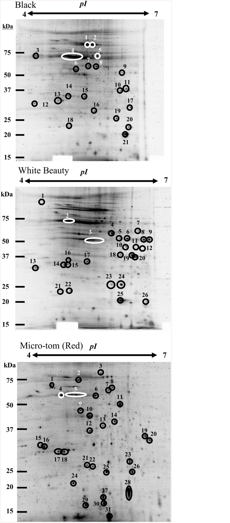
Fig 5. 2D gel electrophoresis of chromoplast proteins in fruit cells of ‘Micro-Tom’ (Red), ‘Black’ and ‘White Beauty’.

Using an 11 cm IPG DryStrip (pH4-7), 100 μg of solubilized proteins were separated. Two-dimensional gel electrophoresis was performed with 12% poly-Acrylamide gel (16 cm x 16 cm). Following electrophoresis, the gel was stained with Flamingo Fluorescent Gel Stain (BIO-RAD). A list of proteins numbered in Fig 5 can be found in Table 1.
