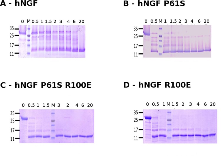
Fig 1. Kinetics of proteolytic cleavage of proNGF WT and mutants.
Representative SDS-PAGE of hproNGF WT (panel A), hproNGF P61S (panel B), hproNGF P61SR100E (panel C), hproNGF R100E (panel D) digested by trypsin. The lanes correspond to aliquots of the reaction mixtures, taken at time 0 (immediately after trypsin addition) and after 0.5, 1, 1.5, 2, 3, 4, 6, 20 hrs. The loading position of the molecular weight marker is indicated by M.

