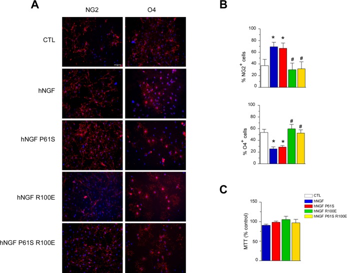
Fig 6. hNGF WT and mutants bioactivity on in vitro differentiation of purified primary rat OPCs.
Panel A: Immunolabelling of primary purified OPC cultures grown for 24 hours in control conditions or in the presence of hNGF WT or its mutants (R100E, P61S, P61S R100E; all compounds are 150 ng/ml). Cell nuclei are marked with DAPI (blue). Scale bar: 50 μm. hNGF increases the percentage of undifferentiated NG2+ cells (left panels: in red) and decreases the percentage of O4+ pre-oligodendrocytes (right panels: in red), indicating that NGF inhibits OPC differentiation. The same effect is observed when cells were growth in the presence of the hNGF mutant P61S but not of R100E nor P61S R100E. Panel B. Quantification of the percentage NG2+ (upper panel) and O4+ (lower panel) OPCs in all different experimental conditions. Three coverslips per experiment were performed in each experimental group. Ten random microscopic fields (20x) per coverslip were evaluated. Experiments were run in triplicate. *P<0.05 vs CTL, #P<0.05 vs hNGF, One-way ANOVA followed by Newman-Keuls post-test. Panel C. None of the compounds tested (all 150 ng/ml; 24 hours exposure) induced toxicity in rat OPC cultures.

