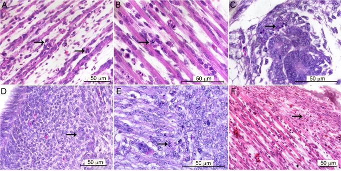
Fig 1. Brightfield microscopy of tissues of Gallus gallus domesticus 72 hpi with yellow fever 17DD virus.
(A) Apoptotic bodies in skeletal muscular tissue; (B) detail of karyorrhexis in muscular bundles; (C) apoptotic bodies in tubular epithelium; (D) apoptotic bodies in muscular region of gizzard; (E) detail of apoptotic bodies in the muscular layer of the gizzard; (F) apoptotic bodies in fibroblastoid cells of perichondrium. Apoptotic nuclei are indicated by black arrows (→). Hematoxylin and Eosin stain.

