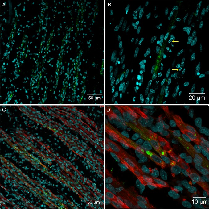
Fig 2. Immunofluorescence in skeletal muscle tissue of chicken embryos at 72 hpi with 17DD virus.
Polyclonal antibodies directed against the yellow fever virus were used to immunostain virus proteins in: (A) skeletal muscle cell bundles; (B) skeletal muscle cells showing perinuclear thickening and presenting an intense labeling in sarcoplasmic reticulum following the striations of the cytoskeleton–yellow arrows (→) show pyknosis and karyorrhexis close to infected cells; and (C) skeletal muscle cells evidenced by desmin antibody and showing the virus infection of the muscular bundles. (D) Detail of the infected muscular bundles, showing the perinuclear positivity together with striations. Yellow fever virus staining in green, nuclei stained with DAPI in blue and desmin in red.

