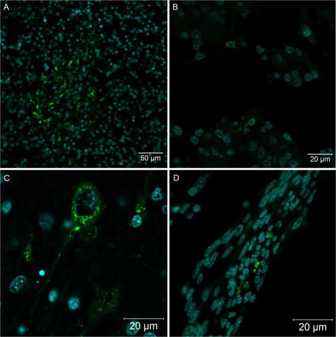
Fig 4. Nervous system of Gallus gallus domesticus at 72 hpi with yellow fever 17DD virus.
(A) Brain section presenting some infected neurons and glial cells; (B) spinal cord infected neurons; (C) one neuron of the brain showing perinuclear thickening and vesicles dispersed throughout the cytoplasm; (D) infected fibroblastoid cells along the meninges. Yellow fever virus protein detection in green and nuclei stained with DAPI in blue.

