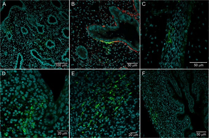
Fig 6. Fibroblastoid cells in different sites of Gallus gallus domesticus at 72 hpi with 17DD virus.
(A) Parenchyma lung cells surrounding the parabronchi epithelium; (B) detail of parenchymal positive cells with desmin expression; (C) infected fibroblastoid cells along the perichondrium; (D) infected cell cluster in subepithelial connective tissue; (E) infected cells in the muscular layer of the gizzard; (F) infected cells in the muscular layer of the yolk stalk. Yellow fever virus proteins immunostained in green, nuclei stained with DAPI in blue and desmin in red.

