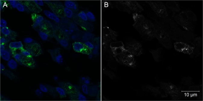
Fig 7. Intracellular aspects of muscle cells of Gallus gallus at 72 hpi with 17DD virus.
(A) Skeletal muscle cells detected by confocal microscopic analysis; (B) Airyscan super-resolution microscopy of the same field of view in a 0.16 μm optical slice showing a vesicular pattern of virus protein expression. Yellow fever virus proteins detected in green (A) or in white (B), nuclei stained with DAPI in blue (A).

