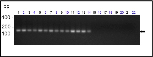
Fig 9. Detection of viral genomic RNA in YF 17DD-infected chicken embryos.

The amplicons generated by Nested-PCR were analyzed by 2% agarose gel electrophoresis. The lanes correspond to the following specimens: (1) and (2)—head; (3) and (4)—legs; (5) and (6)—wings; from (7) to (14)—trunks; (15) and (16)—vitelline membrane; (17) and (18)—chorioallantoic membrane; from (19) to (22)—negative control (water-inoculated animals). Even-numbered lanes indicate samples submitted to amplification of genomic RNA whereas odd-numbered lanes indicate samples submitted to amplification of the replicative intermediate RNA. The molecular length markers are indicated on the left of the figure. The black arrow indicates the 156bp amplicon obtained from the amplification of YF 17D RNA.
