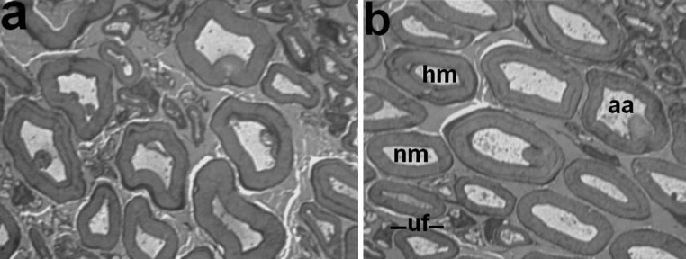
Fig. 3.

Fragments of histological transverse semithin sections of the tibial nerve in dogs with tibial fracture at 37 days of the consolidation phase. Methylene blue-basic fuchsin staining. a Majority of nerve fibres with myelin decompaction and axonal atrophy, b various nerve fibres: nm normally myelinated, hm hypermyelinated, aa severe axonal atrophy, uf profiles of unmyelinated nerve fibres containing nuclei. Instrumental magnification 1250×
