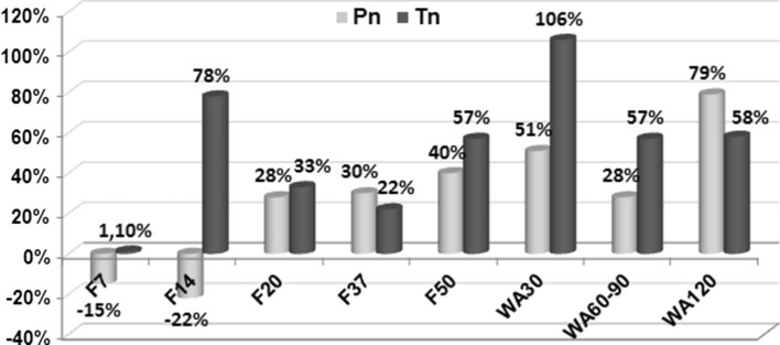
Fig. 4.
Endoneural vessel quantification. Per cents of differences of endoneural vessel densities between nerves of intact (i) and experimental (e) animals (NAmv-i − NAmv-e/NAmv-i × 100 %) in peroneal (Pn) and tibial (Tn) nerves at various time-points of experiment (F7–F50—days of consolidation phase; WA30–WA120—days after the fixator removal)

