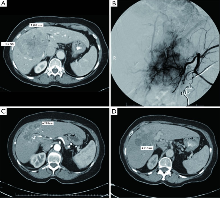
Figure 1.
Large 9 cm liver tumor with corresponding hepatic angiogram showing a highly vascular tumor (A) and coiling of a collateral blood vessel (B). CT scan patient 6 and 12 months after Y-90 radioembolization (C,D). This patient was considered to have a partial response, and later developed progression of disease. CT, computed tomography; Y-90, Yttrium-90.

