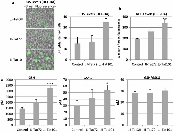
Fig. 3.

Intracellular ROS generation and glutathione levels in Jurkat-Tat101 cells. a Microscopy analysis of intracellular ROS levels measured by DCF-DA staining method. Representative fields of living Jurkat-Tat72, Jurkat-Tat101 and control cells are shown. Acquisition conditions remained the same for each cellular type. The graph shows the number of cells with saturated signal for green laser, from three independent experiments. b Cytometry analysis of DCF-DA stained cells. Graph shows G-mean of green fluorescence intensity of the living cell population from three independent experiments. c Intracellular concentration of reduced (GSH), oxidized (GSSG) and ratio of total glutathione (GSH/GSSG) were measured in Jurkat-Tat72, Jurkat-Tat101 and control cells. Data shown are media and SEM from at least three independent experiments. Kruskal–Wallis test with Dunn’s Multiple Comparison post hoc analysis was performed for statistical analysis (*p < 0.05 and ***p < 0.001 vs control)
