Fig. 2.
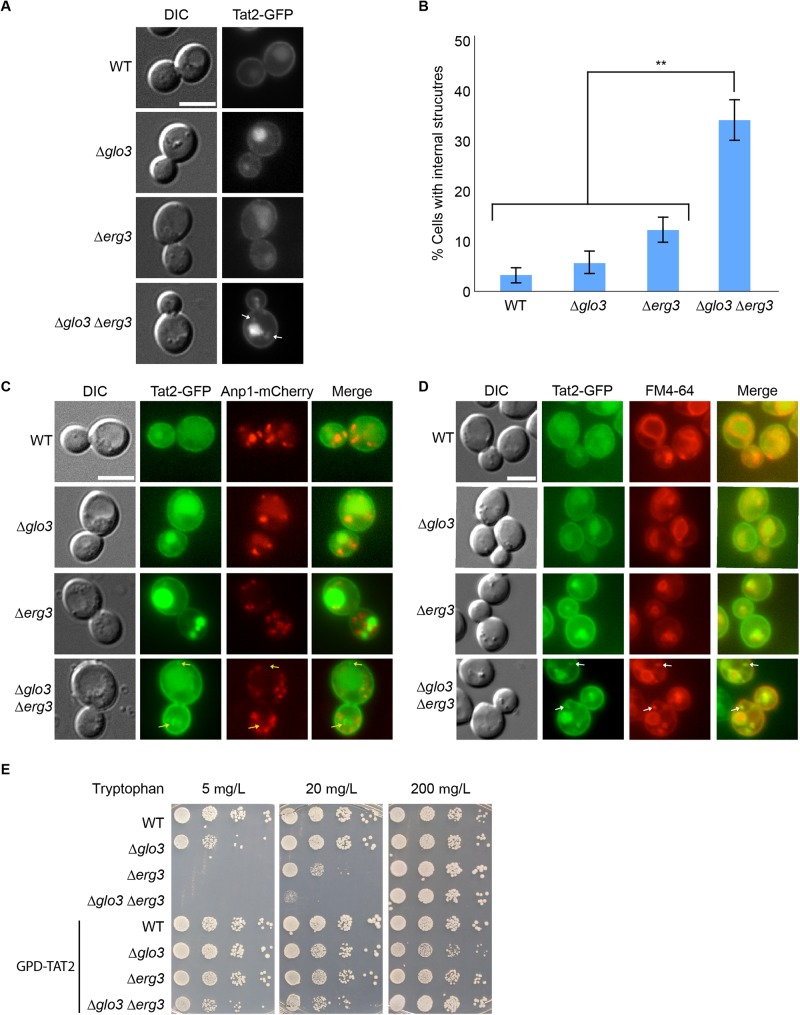
The localization of the tryptophan permease Tat2 is impaired in Δglo3Δerg3 cells. (A) Tat2 accumulates in intracellular foci in Δglo3Δerg3 cells. The localization of Tat2-GFP was assessed in early- to mid-log phase growing cells of different strains. (B) Quantification of A. The data of at least three independent experiments in which≥100 cells were counted per strain are displayed. Error bars represent standard deviation. The p-value corresponds to<0.01. (C) Tat2 does not accumulate in the Golgi. Double labeling of Tat2-GFP and the Golgi marker Anp1-mCherry. Arrows point to non-overlapping signals. (D) Tat2 accumulates in endocytic compartments. Double staining of Tat2-GFP and the lipophilic dye FM4-64, marking endocytic compartments. Arrows point to overlapping signals. (E) Overexpression of Tat2 rescues the growth defect of Δerg3 and Δglo3Δerg3 cells on low tryptophan plates. Drop assay of indicated yeast strains on selective media containing different concentration of tryptophan; 200 mg/l being the standard concentration. The scale bars in A, C and D represent 5 µm.

