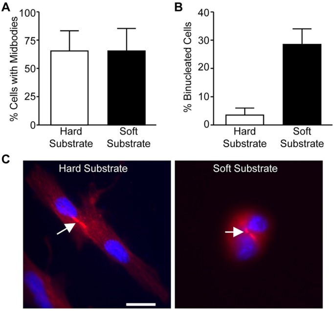
Fig. 5.

Mitotic HDFs form midbodies on both hard and soft substrates. Mitotic HDFs were collected by the shake-off method and replated onto fibronectin-coated hard and soft hydrogels in DMEM with 10% FBS and incubated at 37°C for 1.5 h and 3 h. The cells were fixed and stained for α-tubulin (red) and nuclei (blue). (A,B) Plotted is the percentage of cells with midbodies at 1.5 h (A) and that are binucleated at 3 h (B). Data is reported as the mean±the distribution from two independent experiments in which more than fifty cells were counted per condition for each experiment. (C) Representative images of cells at 1.5 h on hard and soft substrates are shown. Bar, 25 µm. Arrows point to the intercellular bridge/midbody.
