Fig. 3.
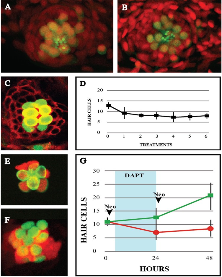
Hair-cell regeneration after recurrent damage. (A-B) Confocal images of a Tg[ET(krt4:EGFP)sqet4] (green) larval neuromast labeled with DAPI (red) showing hair cells (A) before neomycin treatment and (B) 72 h after the 6th treatment. (C) Image of a Tg[ET(krt4:EGFP)sqet4] (green) larval neuromast counterstained for cellular membranes (red) 24 hpt, with 8 hair cells. (D) Graph depicting the number of hair cells per neuromast 24 h after each neomycin treatment, over the course of 6 consecutive treatments. (E-F) A neuromast from a neomycin-treated larva Tg[Atoh1a:dTomato (red) ; ET(krt4:EGFP)sqet4 (green)] without (E), and with Notch inhibition with DAPT (F), showing more numerous hair cells and stronger and broader Atho1a expression. (G) Graph showing number of hair cells per neuromast 24 h after two neomycin treatments with (green) and without (red) inhibition of Notch. DAPT incubation period is shadowed in blue. Results are mean±s.d. Time points: 0 h N=5 neuromasts (5 animals), 24 h N=8 neuromasts (8 animals), 48 h N=4 neuromasts (4 animals).

