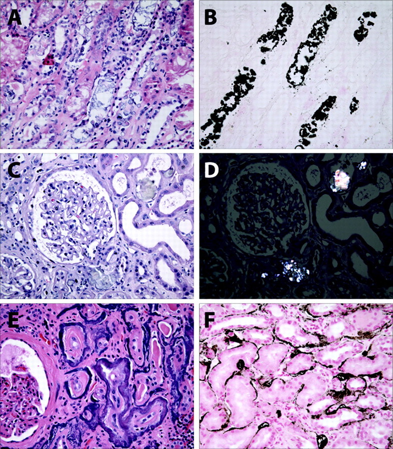
Figure 2.

Renal pathology in nephrocalcinosis. (A) Hematoxylin and eosin-stained biopsy from a patient with phosphate nephropathy. Note the evidence of acute tubular injury with simplified epithelial cell brush borders and necrotic luminal cell debris. (B) von Kossa stain of the same biopsy specimen reveals abundant intraluminal calcium crystals. (C) Hematoxylin and eosin-stained glomerulus from a patient with acute kidney injury and inflammatory bowel disease. (D) On polarized light, the calcium oxalate crystals from the same section are positively birefringent. (E) Hematoxylin and eosin-stained section from a patient with advanced malignancy with bony metastases and hypercalcemia. Note the thickening of tubular basement membranes. (F) von Kossa stain from the same biopsy reveals typical findings in metastatic calcification with punctate and linear tubular basement calcium phosphate crystals.
