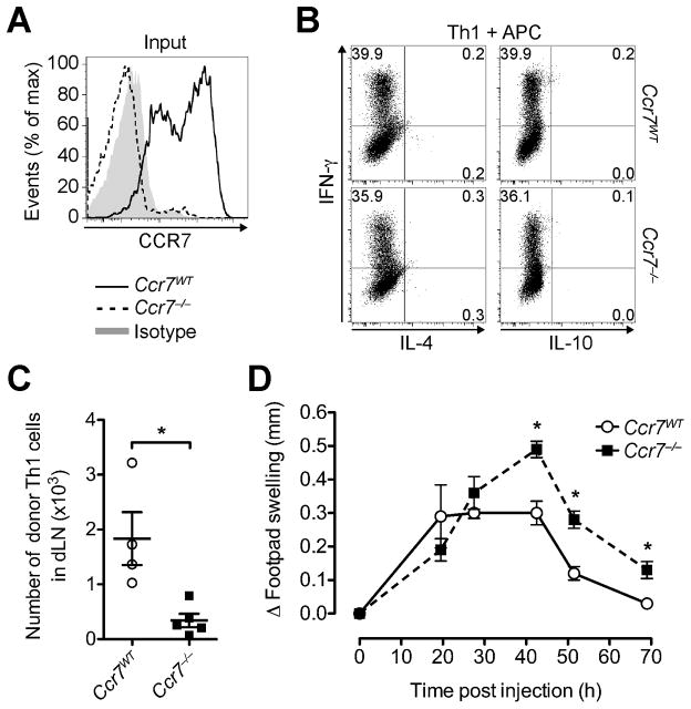
FIGURE 3. Lack of CCR7 impairs effector T cell egress and enhances local inflammation.
Ccr7WT and Ccr7−/− (CD45.2+) OTII Th1 cells were separately co-incubated with OVA-pulsed DCs, before subcutaneous injection into the footpads of two groups of CD45.1+ congenic WT recipients. (A) Representative CCR7 staining of Th1 cells prior to injection. (B) Intracellular cytokine profile of Ccr7WT OTII and Ccr7−/− OTII Th1 cells prior to injection after stimulation with OVA-pulsed APCs. (C) 70 h after transfer, recovered donor Th1 cells were quantified in the dLN. (D) Footpad swelling induced by Ccr7WT and Ccr7−/− OTII Th1 cells was measured over time. Data points depict individual recipients (C) and/or the mean ± SEM of each group (C and D). One experiment of ≥ 2 with similar results using 5 recipients per group. *P < 0.05.

