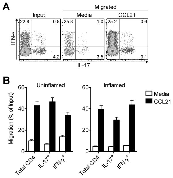
FIGURE 4. Inflammation exiting Th1 and Th17 cells chemotax to CCR7 ligand.
Chronic skin inflammation was induced in sheep by subcutaneous injection of CFA. ≥ 3 weeks later afferent lymph vessels draining the inflamed and uninflamed control skin were cannulated and skin-egressing T cells collected. Chemotaxis of lymph-borne CD4+ T cells toward CCL21 was tested ex vivo in a Transwell chemotaxis assay. (A) Representative intracellular cytokine staining of inflammation-draining CD4+ T cells in input and migrated populations after polyclonal stimulation. (B) Chemotaxis of CD4+ T cell subsets. Bars indicate the mean ± SD for triplicate wells for one representative animal of ≥ 3 analyzed for each condition.

