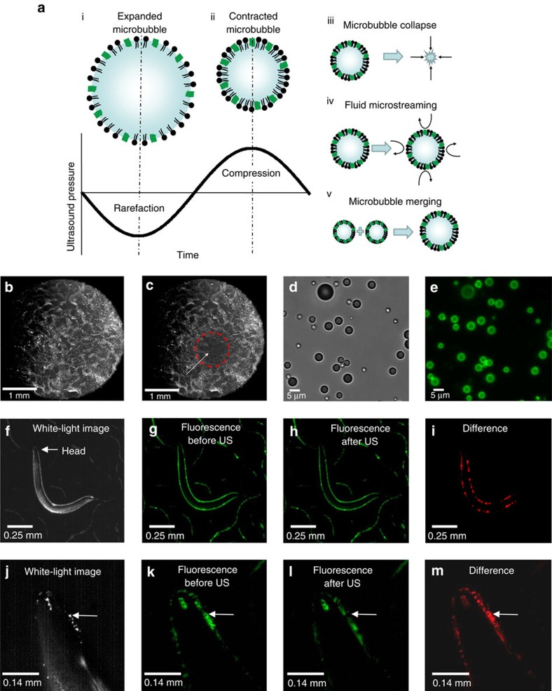
Figure 3. Ultrasound−microbubble interaction with C. elegans.
(a) The microbubble (i) expands and (ii) contracts in size with the rarefaction (low-pressure) and compression (high-pressure) phases of the ultrasound pressure wave. This oscillation behaviour occurs at the frequency of the driving ultrasound, resulting in a variety of behaviours including (iii) microbubble collapse, (iv) fluid microstreaming and (v) merging of microbubbles. These microbubble behaviours create mechanical distortions that can propagate through the agar and the body of the animal. (b) Microbubbles are uniformly distributed on an agar surface and appear white. (c) Ultrasound stimulus (10 pulses lasting 10 ms each with a duty cycle of 1 Hz, 2.25 MHz with a peak negative pressure of 0.9 MPa) activates and destroys the microbubbles in the ultrasound focal zone of 1 mm diameter (white arrow). Microbubbles outside this focal zone (denoted by the red circle) appear undisturbed. Microbubbles were labelled with DiO, and (d) brightfield and (e) fluorescent images are shown. A whole-animal view showing (f) brightfield, (g) fluorescence before and (h) after ultrasound stimulus and finally, (i) the difference in red showing the locations of microbubble-induced mechanical deformations. A magnified view of the animal's head in (j) brightfield, (k) before and (l) after ultrasound stimuli (US) and (m) the difference in red showing the locations of microbubble-induced mechanical deformations. The white arrow points to a large microbubble that is destroyed upon ultrasound stimulation.

