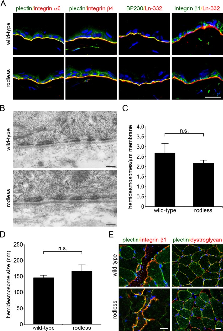
FIGURE 3:
Rodless plectin mice show normal plectin localization and HD organization. (A) Frozen sections of the skin of wild-type and rodless plectin mice were double labeled for the indicated proteins. Nuclei were counterstained with DAPI. Scale bar, 20 μm. (B) Electron microscopy images of the skin of wild-type and rodless plectin mice showing hemidesmosomal structures. Scale bars, 200 nm. (C, D) Quantification of the results shown in B. The number of HDs per length unit of plasma membrane (C) and size of HDs (D) was quantified in two wild-type and two rodless plectin mice. Values represent the mean and SD. n.s., not significant. (E) Frozen sections of skeletal muscle of wild-type and rodless plectin mice were stained for plectin and integrin β1 or dystroglycan. Nuclei were counterstained with DAPI. Scale bars, 20 μm.

