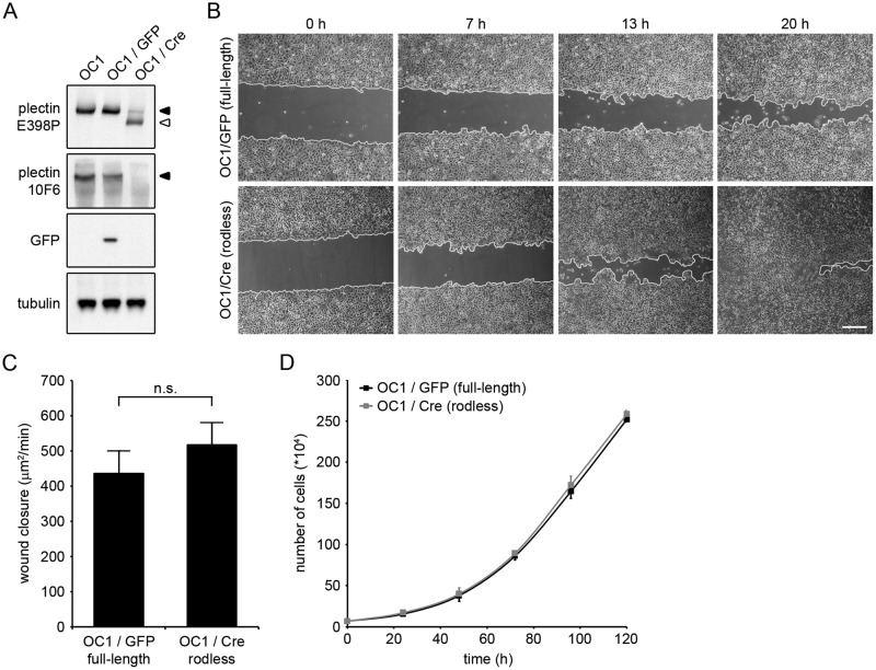
FIGURE 5:
Keratinocytes expressing rodless plectin show a slight but consistent increase in cell migration. (A) OC1 Plecfl(Ex31),fl(Ex31) keratinocytes (OC1) were infected with adenoviruses expressing either GFP (OC1/GFP) or Cre-recombinase (OC1/Cre). Lysates of OC1, OC1/GFP and OC1/Cre were analyzed by Western blot for expression of the indicated proteins. The 10F6 and E398P plectin antibodies recognize epitopes in the rod domain and C-terminus, respectively. Tubulin levels served as a loading control. (B) Phase contrast images of OC1/GFP and OC1/Cre at 0, 7, 13, and 20 h after wounding. Wound edges are marked in white. Scale bar, 50 μm. (C) Rate of wound closure in OC1/GFP and OC1/Cre cells. Values represent the mean ± SEM of three independent experiments. n.s., not significant. (D) Proliferation of OC1/GFP and OC1/Cre cells. Error bars indicate the SD over two independent experiments.

