FIGURE 1:
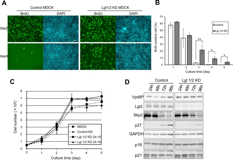
Lgl is involved in suppression of proliferation in confluent epithelial cells. (A) Control MDCK and Lgl1/2 KD MDCK cells were seeded and cultured for the indicated times. BrdU was added 3 h before fixation, and BrdU-positive cells were visualized by immunofluorescence. Scale bar, 50 μm. (B) The ratio of BrdU-positive cells to total cells was determined, and averages of three independent experiments are plotted. Error bar indicates ±SD. Single and double asterisks denote significant differences p < 0.05 and p < 0.01, respectively, by Student's t test. (C) A total of 5 × 10⁴ normal MDCK, control MDCK, and two Lgl1/2 KD MDCK cell clones was seeded in 12-well Transwell plates and counted using a hemocytometer. Error bars indicate ±SD of three independent experiments. Note that Lgl1/2 KD MDCK cells grew to a significantly higher saturation density than normal MDCK or control MDCK cells (p < 0.05, Student's t test between all four combinations at day 4). (D) Control MDCK and Lgl1/2 KD MDCK cells were seeded and cultured until the indicated times. Then levels of cell cycle inhibitors and Skp2 were monitored. Cell density–dependent induction of p27 and suppression of Skp2 were attenuated in Lgl1/2 KD MDCK cells.

