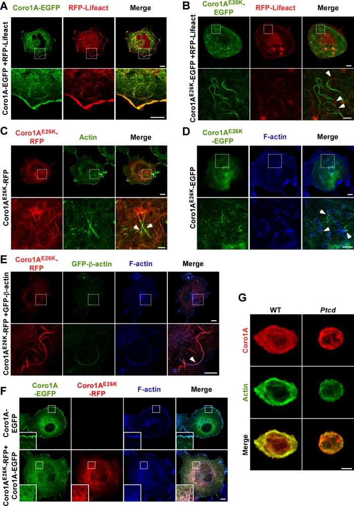
FIGURE 3:
Coro1AE26K-nucleated filaments contain F-actin. (A, B) Confocal images of COS1 cells coexpressing RFP-LifeAct (A and B, red signals) in the presence of either Coro1A-EGFP (A, green signals) or Coro1AE26K-EGFP (B, green signals). In A and B, enlarged images of indicated cell areas (white open squares) are shown at the bottom. Potential areas of colocalization between RFP-LifeAct and Coro1A proteins had to be seen in yellow. Some of the filament areas found positive for RFP-LifeAct epifluorescence are indicated by arrowheads (B). Scale bars, 10 μm (top), 5 μm (bottom); same scales in C–F. (C) Confocal images of Coro1AE26K-RFP–expressing COS1 cells stained with antibodies to actin. Ectopic Coro1A and endogenous actin proteins are seen in red and green, respectively. Areas of colocalization had to be seen in yellow in the right column. Enlargements of indicated cell areas (white open squares) are shown in the appropriate bottom images. Some of the filament areas found positive for actin immunoreactivity are indicated by arrows. (D) Coro1AE26K-EGFP–expressing cells were stained with Alexa Fluor 635–phalloidin and analyzed by confocal microscopy. Enlargements of the indicated cell areas (top, white open squares) are shown in the appropriate bottom images. Coro1AE26K-EGFP and F-actin signals are seen in green and blue, respectively. Areas of colocalization had to be seen in white. Arrowheads show segments of filaments that are phalloidin-positive (bottom). (E) COS1 cells coexpressing Coro1AE26K-RFP and GFP–β-actin were stained with Alexa Fluor 635–phalloidin and analyzed by confocal microscopy. Enlargements of the indicated cell areas (top, white open squares) are shown in the appropriate bottom images. Epifluorescence signals for Coro1A and β-actin are shown in red and green color, respectively. F-actin signals are seen in blue. Areas of colocalization for those two proteins are seen in purple. Arrowhead shows a filament segment that displays significant colocalization between these two proteins (bottom). (F) COS1 cells expressing the indicated combination of wild-type and Coro1AE26K proteins (top) were stained with Alexa Fluor 635–labeled phalloidin and analyzed by confocal microscopy. RFP and EGFP epifluorescence signals are seen in red and green, respectively. F-actin–rich areas are in blue. Areas of colocalization between RFP- and EGFP-tagged proteins are in yellow. Colocalization areas for RFP, EGFP, and F-actin had to be seen in white. Insets, enlarged images of the indicated cell areas (white open squares). (G) Confocal Z-stack 3D reconstructions of thymocytes isolated from mice of indicated genotypes (top) and stained with antibodies to Coro1A (top and bottom; red signals) and to actin (middle and bottom; green signals). Potential areas of colocalization between Coro1A and actin had to be seen in yellow (bottom). Scale bar, 2.5 μm.

