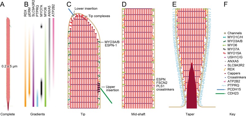
FIGURE 2:
Model chick vestibular stereocilium. Scale model of stereocilium showing selected molecules; these molecules are drawn in at the approximate density for each as determined by mass spectrometry experiments (Shin et al., 2013). (A) Complete stereocilium at low magnification showing dimensions. (B) Selected molecular gradients in the stereocilia. See Shin et al. (2013) for details. (C) High-magnification view of the tip region of the stereocilium. The two insertions of the tip link are highlighted; the lower insertion includes transduction channels, and the upper insertion has USH1C, USH1G, and MYO7A. In addition, the MYO15A tip complex is indicated; DFNB31, EPS8, and perhaps EPS8L2 are part of this complex. MYO3A and MYO3B are found, along with their cargo ESPN-1, in the tip region as well. (D) The stereocilia shaft is made of parallel actin filaments cross-linked by a variety of proteins, most prominently ESPN, FSCN2, and PLS1. Major components of the shaft include the calcium pump ATP2B2, the membrane-associated ANXA5, the actin-to-membrane connector radixin (RDX), and SLC9A3R2, a ligand for RDX. RDX with the activating phosphorylation is found above the taper region. (E) The taper region. Most actin filaments terminate at the membrane, but a few project through into the soma as the rootlet (density). The lipid phosphatase PTPRQ is concentrated in the taper region, as is unactivated RDX. (F) Key to molecules included in the diagram.

