FIGURE 1:
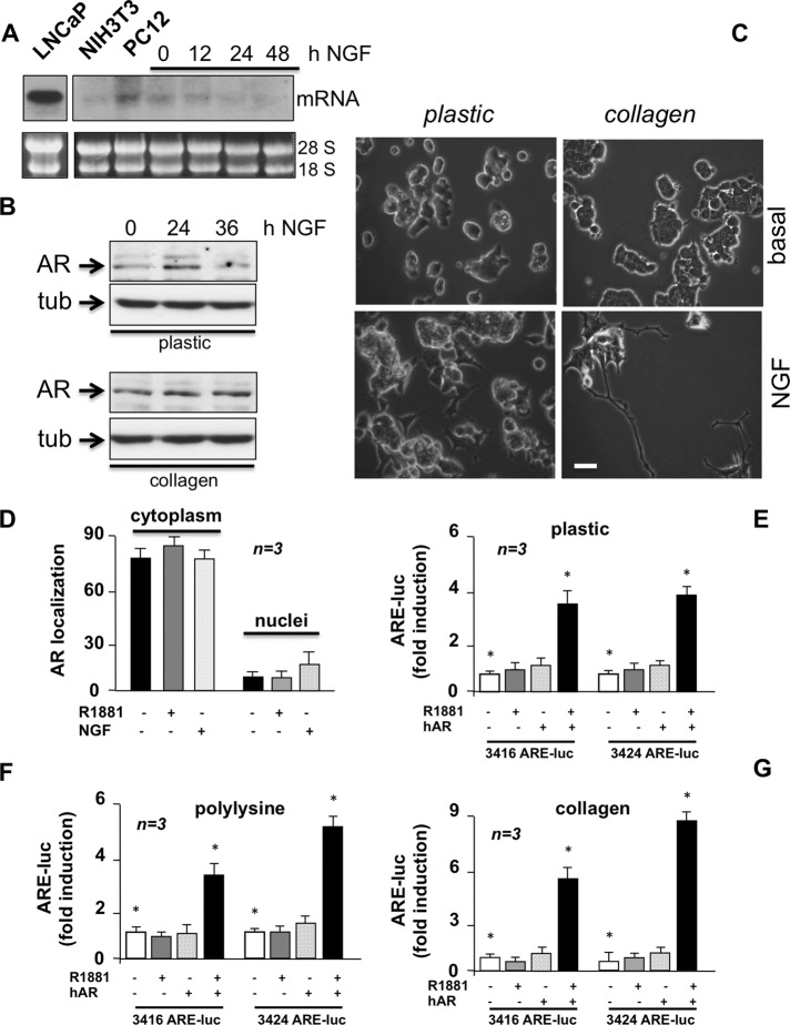
PC12 cells harbor extranuclear classic AR. (A) Growing LNCaP, NIH3T3, and PC12 cells on plastic were used. PC12 cells were made quiescent for 12 h and then stimulated for the indicated times with NGF (100 ng/ml). mRNA was extracted and used for Northern blot of AR mRNA (top). The loading mRNA control (28S and 18S) was analyzed (bottom). (B) Plastic- or collagen-plated PC12 cells were made quiescent, then left untreated or treated for the indicated times with NGF (100 ng/ml). AR expression was detected by Western blot of lysate proteins. The filters were reprobed with anti-tubulin antibody as a loading control. (C) Plastic- or collagen-plated PC12 cells were made quiescent and then challenged for 24 h with NGF (100 ng/ml). Contrast phase images are representative of three different experiments, each performed in duplicate. Bar, 5 μM. (D) PC12 cells on polylysine-coated coverslips were made quiescent and then left untreated or treated for 1 h with R1881 (10 nM) or NGF (100 ng/ml). Cells were stained by IF for AR. Total nuclei were stained with Hoechst, and AR intracellular localization was analyzed. Data are represented as percentage of cells showing exclusively nuclear or cytoplasm AR staining. Data from at least 500 cells from each independent experiment were scored and are graphically presented. Means and SEM are shown; n represents the number of experiments. Growing PC12 cells plated on plastic (E), polylysine (F), or collagen (G) were transfected with either 3416 or 3424 ARE-Luc constructs with or without hAR-expressing plasmid. Cells were made quiescent and left unstimulated or stimulated for 18 h with 10 nM R1881. Luciferase activity was assayed, normalized using β-galactosidase as an internal control, and expressed as fold induction. Data from several independent experiments were analyzed. Means and SEM are shown; n represents the number of experiments. The statistical significance of results was also evaluated by paired t test in all the experiments (*p < 0.05). The difference in ARE-Luc induction between cells challenged with 10 nM R1881 and unstimulated cells was significant (*p < 0.05) only in cells cotransfected with hAR and 3416 ARE-Luc or 3424 ARE-Luc.

