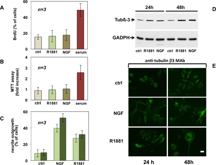
FIGURE 3:
Androgen does not stimulate proliferation but triggers differentiation of PC12 cells. Quiescent PC12 cells were used. (A) Cells on polylysine-coated coverslips were left unstimulated or stimulated for 24 h with R1881 (10 nM), NGF (100 ng/ml), or serum (at 20%). Cells were pulsed for 4 h with 100 μM BrdU (Sigma-Aldrich). BrdU incorporation was analyzed by IF and expressed as percentage of total cells. (B) Plastic-plated cells were left unstimulated or stimulated for 24 h with R1881 (10 nM), NGF (100 ng/ml), or serum (at 20%). After 24 h, MTT assay was performed. (C) Cells on polylysine-coated coverslips were left unstimulated or stimulated for 24 h (light green) or 48 h (dark green) with R1881 (10 nM) or NGF (100 ng/ml). Neurite outgrowth was analyzed by contrast phase microscopy and expressed as percentage of total cells. In A–C, data derive from several independent experiments. Means and SEM are shown. (D) Plastic-plated cells were left untreated (ctrl) or challenged for the indicated times with R1881 (10 nM) or NGF (100 ng/ml). Expression of tubulin β-III (tub β-III) was analyzed by Western blot of lysate proteins. Filters were reprobed with anti-GADPH as a loading control. (E) Cells on polylysine-coated coverslips were left untreated or treated for the indicated times with R1881 (10 nM) or NGF (100 ng/ml). Cells were analyzed by IF for tubulin β-III. Bar, 10 μM.

