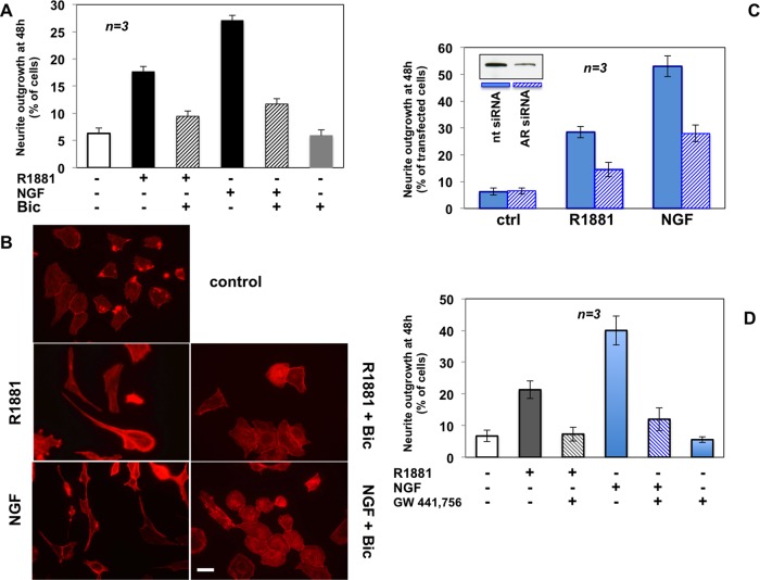
FIGURE 4:
Neurite outgrowth in PC12 cells stimulated by R1881 or NGF: functional cross-talk between AR and TrkA. PC12 cells were used. (A, B) Polylysine-plated cells were made quiescent and then left untreated or treated for 48 h with 10 nM R1881 or 100 ng/ml NGF in the absence or presence of 10 μM bicalutamide. (A) Neurite outgrowth expressed as percentage of total cells. (B) Actin stained using Texas red–phalloidin and analyzed by IF. Images are representative of two independent experiments. Bar, 10 μM. (C) Growing cells on polylysine-coated coverslips were transfected with AR or nt siRNA. Purified pEGFP (Amaxa) plasmid was included to help identification of transfected cells. Cells were made quiescent and then left unchallenged or challenged with R1881 or NGF. GFP-expressing cells were visualized by fluorescence microscopy and their neurite outgrowth analyzed by contrast phase microscopy and expressed as percentage of transfected cells. Several independent coverslips were analyzed, and data from at least 200 scored cells for each coverslip were collected and are graphically shown. Inset in C shows AR protein levels detected by Western blot of lysate proteins from PC12 cells transfected with nt or AR siRNA. About 30% of AR protein was still detected upon AR siRNA, as evaluated by ImageJ software (National Institutes of Health, Bethesda, MD). (D) Quiescent cells on polylysine were left untreated or treated for 48 h with R1881 or NGF in the absence or presence of the TrkA inhibitor GW441776 (1 μM). Neurite outgrowth was evaluated and expressed as percentage of total cells. In A, C, and D, means and SEM are shown.

